
Featured Instrumentation
Vit-Buckle Academy and the Vit-Buckle Society proudly recognize and thank our industry sponsors for their generous support.
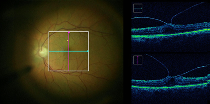
ZEISS RESCAN 700
With innovative technologies such as RESCAN® 700 integrated intraoperative OCT (io-OCT) from ZEISS, the OPMI LUMERA 700 from ZEISS provides superb clarity and clinical insights when performing retina surgery procedures. The iiOCT adds a real-time third dimension to visualization capabilities for viewing transparent structures of the eye.
RESCAN in your OR
Audina Berrocal, MD
Professor of Clinical Ophthalmology
Medical Director of the Retinopathy of Permaturity Service
Bascom Palmer Eye Institute – Miami, FL
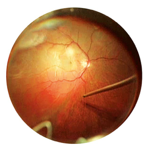
ZEISS RESIGHT Fundus Viewing System
Achieve optimum visualization and performance with RESIGHT® from ZEISS. The non-contact fundus viewing system provides a clear, detailed view of the retina. RESIGHT incorporates varioscope optics to keep you in focus on the retina without moving the microscope and allows you to seamlessly switch between wide angel and macular lenses.
RESCAN in your OR
Audina Berrocal, MD
Professor of Clinical Ophthalmology
Medical Director of the Retinopathy of Permaturity Service
Bascom Palmer Eye Institute – Miami, FL
ZEISS AngioPlex® OCT Angiography on our CIRRUS™ HD-OCT
The introduction of ZEISS AngioPlex ushers in a new era in retinal care. Now, ultra-clear 3D microvascular visualizations with non-invasive technology can be a routine part of your retina practice.
- New vascular information Depth–resolved visualization of the retinal vasculature. Powered by Optical Micro Angiography (OMAGc) Algorithms that utilize amplitude and phase OCT signal data to deliver the highest-quality ultra-clear 3D angiography images.
- Enhanced workflow FastTrac™ provides live-tracking for motion-artifact-free images. Single-Scan Simplicity ensures ZEISS AngioPlex requires only a single additional OCT scan to generate an ultra-clear 3D OCT angiography image.
- Most powerful OCT platform AngioPlex OCT Angiography is available on the CIRRUS 5000 HD-OCT platform, allowing ophthalmic practices the flexibility to easily integrate vascular imaging with standard OCT diagnostic imaging.