

IMAGE OF THE MONTH: February 2022
6 year old boy with blurred vision after trauma


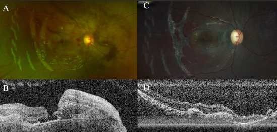
A 6 year old boy presents with blurred vision and pain one week following trauma. Visual acuity is finger counting OD and 20/30 OS.
DIAGNOSIS
Traumatic choroidal rupture with full thickness macular hole after blunt, closed globe injury
DISCUSSION
The differential diagnosis of yellow-white curvilinear or crescent shaped lesions concentric to the optic disc includes lacquer cracks, angioid streaks, and sclopetaria. The diagnosis of choroidal rupture was made based on a history of ocular trauma, characteristic appearance on examination, as well as visualization of the discontinuity in the RPE-Bruch’s membrane complex on OCT. A full thickness macular hole was also best appreciated on OCT. The patient was evaluated in the retina clinic one month after initial presentation. The VA improved to 5’/200E OD. Large chorioretinal curvilinear scars in the temporal macula with gray-brown coloration and peripapillary subretinal fibrosis were observed on follow-up examination. Optic nerve pallor was also noted. The full thickness macular hole appeared improved from initial OCT examination with only observation. The patient is being monitored for secondary choroidal neovascular membrane following choroidal rupture.
Have an interesting image to share?
Email it to us at vitbuckleimage@gmail.com