

IMAGE OF THE MONTH: November 2021
Yellow spots in the retina

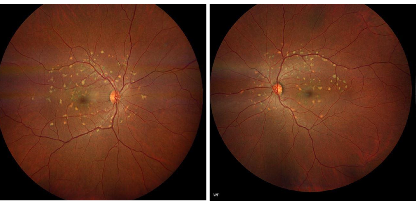
A 45 year old healthy female was referred by her ophthalmologist due to the abnormal fundus exam shown below. She has no family history of eye disease. The patient was asymptomatic and visual acuity was 20/20 OD and 20/20 OS.
DIAGNOSIS
Retina evaluation revealed pisciform flecks in the macula and midperiphery in both eyes, fairly symmetric, with hyperautofluorescence in areas of the flecks.
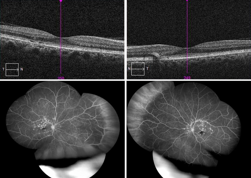
OCT revelaed hyperreflecitivty and irregularity of the retinal pigment epithelium, as well as small linear deposits exnteding into the outer nuclear layer. Fluorescein angiography revealed hyperfluorescent staining of the flecks.
A diagnosis of fundus flavimaculatus was made, and genetic testing subsequently revealed compound heterogenous mutations in ABCA4 – c.5882G>A (pathogenic with low penetrance) and c.1018T>C (likely pathogenic).
Mutations in ABCA4 (ATP-biding cassette-4) are implicated in a variety of retinal conditions, most commonly Stargardt disease and fundus flavimaculatus, but also cone-rod dystrophies anad some forms of retinitis pigmentosa. The severity of the ABCA4 mutation, as well as its penetrance, impact ABCA4 expression levels, which in turn impact the clinical phenotype. Fundus flavimaculatus is considered to be on the milder side of the Stargardt spectrum, with later onset and less visual impairment, than Stargardt. The flecks visible clinically reflect lipofuscin accumulation in the retinal pigment epithelium. The location of the flecks may change over time and does not correlate will to vision. Whereas in Stargardt disease, the flecks tend to be more central and confluent, flecks extend further peripherally and into the midperiphery in fundus flavimaculatus.
Have an interesting image to share?
Email it to us at vitbuckleimage@gmail.com