

IMAGE OF THE MONTH: August 2023
Blurry vision and a bump

Introduction: An adult male in his 30s presents with decreased vision in the left eye.
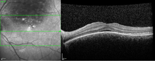
OCT
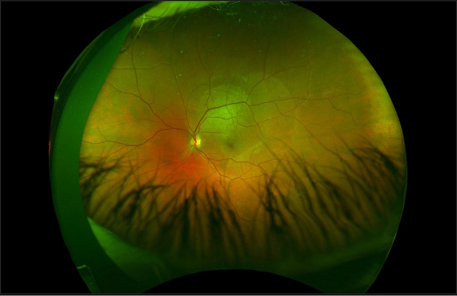
Fundus photo
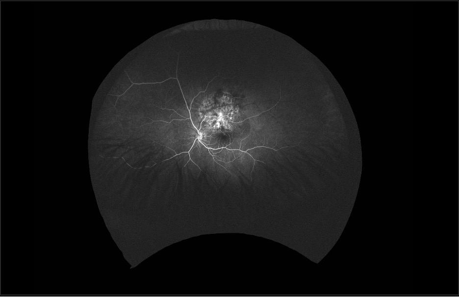
FA (20 seconds)
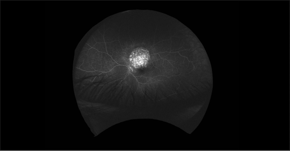
FA (3 minutes)
DIAGNOSIS
Choroidal hemangioma
Choroidal hemangioma is a congenital hamartoma which typically manifests with a unilateral orangish-red lesion, often in the posterior pole. Rarely, a diffuse subtype may be noted, which may be associated with Sturge-Weber syndrome and associated congenital glaucoma. While most circumscribed choroidal hemangiomas are asymptomatic, subretinal fluid (or less commonly, degeneration of retina overlying the lesion) may result in vision decrease, scotoma, or metamorphopsia. Imaging features include elevation with possible overlying subretinal fluid or retinal degeneration on optical coherence tomography. Fluorescein angiography reveals both early and late diffuse hyperfluorescence. B-scan ultrasonograpy classically reveals high internal reflectivity and acoustic solidarity. There may be associated subretinal fluid. The patient described herein developed worsening subfoveal subretinal fluid with vision disturbance and was successfully treated with photodynamic therapy with resolution of fluid and improvement in vision.
Have an interesting image to share?
Email it to us at vitbuckleimage@gmail.com