

IMAGE OF THE MONTH: April 2023
Widespread pigmentation


A 54-year-old woman presented to clinic with 6-month history of intermittent flashes of light in both eyes. Patient denied changes in vision, new floaters, or veils over vision. She denied any family history of ocular conditions. BCVA was 20/20 OD and 20/20-1 OS. The anterior segment was pertinent for trace nuclear and cortical changes in both eyes, otherwise unremarkable.
DIAGNOSIS
Pigmented paravenous retinochoroidal atrophy (PPRCA), OU
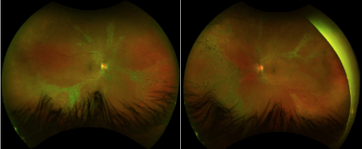
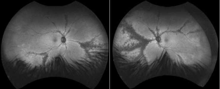
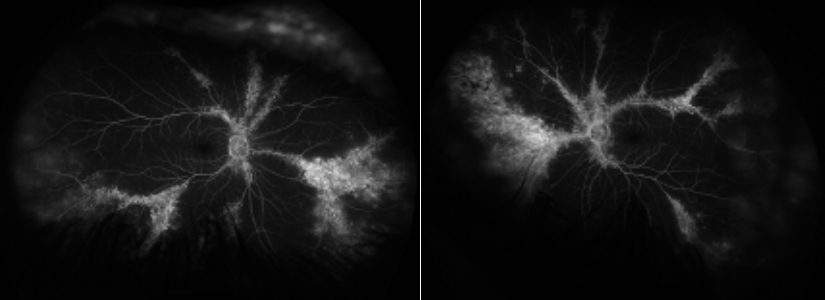
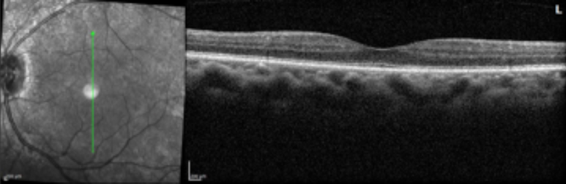
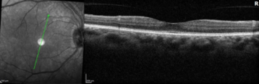
Posterior segment findings were relevant for bilateral areas of localized atrophy and mild hyperpigmentation along retinal veins, extending from the optic nerve head to the periphery. Fundus autofluorescence revealed hypoautofluorescence along these areas. Fundus fluorescein angiography demonstrated hyperfluorescence along the perivenular atrophic areas and minimal blockage in areas of pigment clumping. Optical coherence tomography showed intact retinal laminations with PVD in the right eye and partial PVD in the left eye. The patient was evaluated for tuberculosis and syphilis with negative results. She was diagnosed with pigmented paravenous retinochoroidal atrophy of both eyes.
Pigmented paravenous retinochoroidal atrophy is a retinal degeneration with unclear etiology. Inflammatory, infectious, and genetic causes have been thought to play a role in this retinal degeneration but none of these etiologies have been proven. It is characterized by accumulation of pigment and retinochoroidal atrophy along the veins. Patients are usually asymptomatic. Therefore, most patients are usually diagnosed during routine ophthalmic examinations. Some patients, however, may present with severe vision loss. There is no specific treatment for this rare disorder. This patient has been observed for six years without progression on exam and imaging.
References
Choi WJ, Joo K, Park KH. Pigmented Paravenous Retinochoroidal Atrophy. Korean J Ophthalmol. 2020;34(1):90-91.
Huang HB, Zhang YX. Pigmented paravenous retinochoroidal atrophy (Review). Exp Ther Med. 2014;7(6):1439-1445.
Ramtohul P, Chehaibou I, Bonnin S. Peripheral retinal vascular abnormalities in pigmented paravenous retinochoroidal atrophy. Am J Ophthalmol. 2022;236:e4-e5.
Have an interesting image to share?
Email it to us at vitbuckleimage@gmail.com