

IMAGE OF THE MONTH: September 2022
Blurry vision and red eye for several months

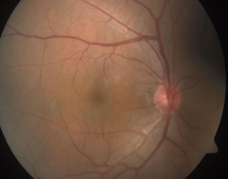

A 64 year-old male was referred by his optometrist for retinal detachment in the left eye. He reported blurry vision and red eye OS for a few months.
DIAGNOSIS
Chorioretinal folds due to orbital mass lesion
DISCUSSION
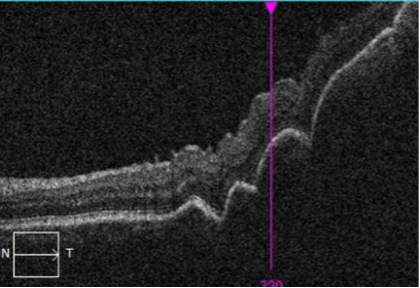
The differential diagnosis of choroidal folds includes: acquired hyperopia, retroorbital/orbital mass/tumor, thyroid eye disease, choroidal tumors, hypotony, inflammation, neovascular membranes, extraocular hardware, papilledema, and idiopathic. Choroidal folds are thought to occur when compressive forces on the choroid, RPE, and Bruch’s membrane exceed the tensile force of choroid due to intraocular pressure. Fundus examination will reveal folding and color banding of the retina/choroid. Fundus autofluorescence and fluorescein angiography reveal alternating bands of hyper and hypo-autofluoresence or fluorescence, respectively, without leakage. Clinical history and additional examination findings will generally identify a causative etiology. In the case above, the patient exhibited dilated and injected scleral/episcleral vessels temporally OS, as well as limitation of adduction and supraduction OS with diplopia on far right or upward gaze. Examination revealed choroidal folds and an apparent mass effect from an orbital lesion. OCT confirmed a choroidal vs. orbital lesion with mass effect on the retina and overlying chorioretinal folds (image below). The patient underwent CT imaging which revealed a ~1x1x1 cm mass lesion in the left orbit, and he was referred to oculoplastics/ocular oncology for further workup and management.
Have an interesting image to share?
Email it to us at vitbuckleimage@gmail.com