

IMAGE OF THE MONTH: April 2022
Abnormal retinal vessels
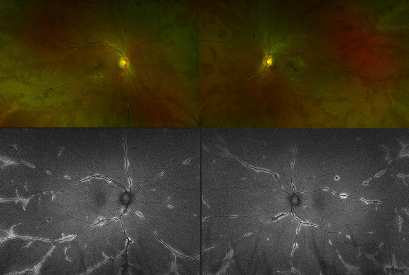
A 29-year-old man referred for abnormal fundus findings seen on routine eye exam. Visual acuity is 20/20 bilaterally and he is asymptomatic. The fundus images and autofluorescence findings are presented below. What is the diagnosis?
DIAGNOSIS
Pigmented paravenous retinochoroidal atrophy
Discussion
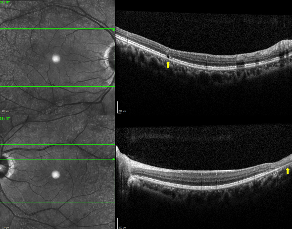
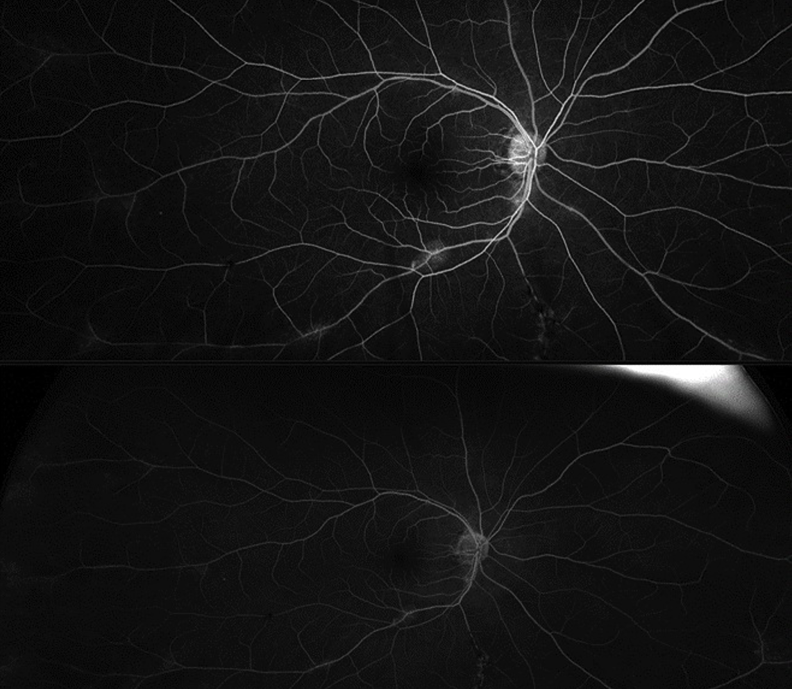
Retinal evaluation revealed perivenular pigmentary changes in both eyes associated with chorioretinal atrophy. The intervening retina was uninvolved. OCT revealed focal areas of retinal pigment epithelium and outer retinal loss along veins (left eye OCT, yellow arrow), with few areas of frank focal outer retinal cavitations clearly distinct from vessel shadowing (right eye OCT, yellow arrow) in these areas in both eyes. Notably, there was no pre-retinal cell or vitreous debris. Inner retinal layers were uninvolved. Fluorescein angiography revealed few areas of early window defects with faint areas staining of chorioretinal scars along veins in both eyes with no significant areas of nonperfusion or active areas of leakage (early 37 seconds and late 5-minute frames right eye shown here). Workup was negative for ESR, CRP, ANCA, ACE, lysozyme, RF, anti-dsDNA, HLA-B27, RPR, FTA-Abs, Quantiferon gold, Lyme, Toxoplasmosis, Coxsackie and COVID serologies. Given the absence of inflammatory findings on exam and imaging, negative work-up, and asymptomatic and stationary nature of the disease, a diagnosis of pigmented paravenous retinochoroidal atrophy (PPRCA) was made. Genetic testing was unrevealing. PPRCA is a rare stationary retinal disease characterized by perivenous bone spicule pigmentation. The etiology is unknown, with most cases occurring sporadically. Familial or hereditary cases have been described, with autosomal dominant inheritance caused by heterozygous mutations in the crumbs analog 1 (CRB1) gene reported in one family, and autosomal dominant inheritance proposed in another case involving amutation in the hexokinase 1 (HK1) gene. Congenital and inflammatory etiologies have also been hypothesized. More cases have been reported in males than females. Patients are generally asymptomatic due to the slow or nonprogressive nature of the disease. There is no treatment for the chorioretinal atrophy. Huang, H. B., & Zhang, Y. X. Pigmented paravenous retinochoroidal atrophy (Review). Exp Ther Med. 2014 Jun;7(6):1439–1445. https://doi.org/10.3892/etm.2014.1648 McKay, G. J., Clarke, S., Davis, J. A., Simpson, D. A., & Silvestri, G. Pigmented paravenous chorioretinal atrophy is associated with a mutation within the crumbs homolog 1 (CRB1) gene. Invest Ophthalmol Vis Sci. 2005;46(1):322–328. https://doi.org/10.1167/iovs.04-0734 Shah SM, Schimmenti LA, Chiang J, Iezzi R. Association of pigmented paravenous retinochoroidal atrophy (PPRCA) with a pathogenic variant in the HK1 gene. Retin Cases Brief Rep. 2020 Nov 4; Epub ahead of print.doi: 10.1097/ICB.0000000000001077.
Have an interesting image to share?
Email it to us at vitbuckleimage@gmail.com