

IMAGE OF THE MONTH: January 2024
Looking Past the Mass


A 50-year-old man presented to clinic with a 2 week history of blurry vision in the right eye assocaited with a pressure sensation along the upper brow. He denied a history of any family history of ocular conditions. BCVA was 20/20 OD, and 20/20 OS. Intraocular pressure was 11mmHg OD and 12mmHg OS. The anterior segment exam was otherwise unremarkable.
Diagnosis
Malignant choroidal melanoma
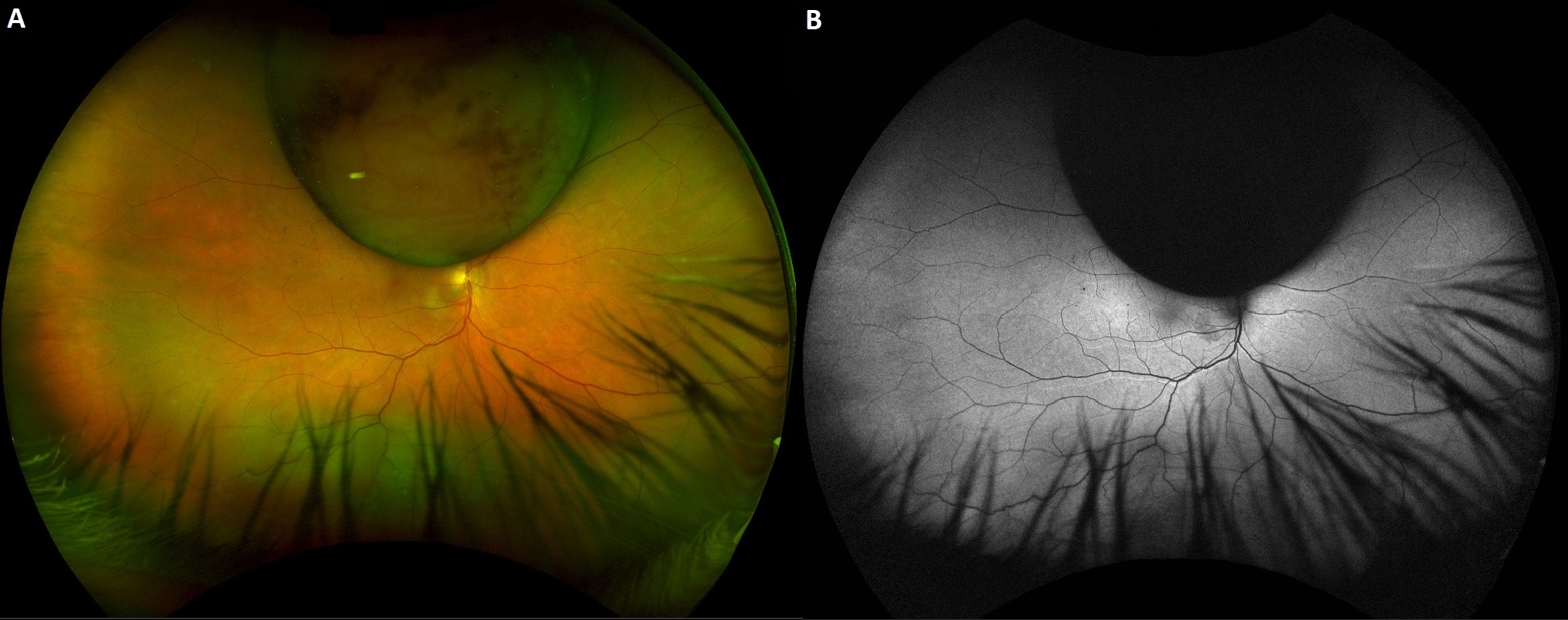
Posterior segment examination was notable for a large mass arising from the superior choroid and an inferior serous retinal detachment (Figure A). The mass measured 11.5mm on B-scan with a mushroom configuration extending through bruchs membrane and into the vitreous cavity. There was an assocaited serous retinal detachment extending from 3 o'clock to 9 o'clock. Fundus autofluorescence (Figure B) revealed hypoautofluorescence in the area of the mass and hyperautoflurescence along the inferior retinal vasculature (retinal printings) secondary to low-integrity retinal attachment (LIRA) or retinal displacement. The patient was referred to an ocular oncologist who confirmed the diagnosis and the patient subsequently underwent I-125 plaque therapy.
Uveal melanoma, remains the most common primary intraocular tumor in adults1 and most commonly occurs unilaterally and in Caucasians. It has a propensity for metastasis to the liver, and a poor prognsis once the tumor has disseminated. The choroid represents the predominant site affected, giving rise to 90% of uveal melanomas. For uncomplicated uveal melanomas, brachytherapy remains the treatment of choice.
Retinal displacement is a common and unwanted outcome following rhegmatogenous retinal detachment repair2. Shiragami et al3 first observed and documented retinal displacement in 2010 through the utilization of fundus autofluorescence (FAF) imaging. In their study, they identified hyperautofluorescent lines positioned alongside and mirroring the shape of retinal vessels. The researchers proposed the term "retinal vessel printings" (RVPs) for these hyperautofluorescent patterns, suggesting that retinal displacement had exposed retinal pigment epithelium (RPE) cells that were previously shielded from light.
References
1. Singh AD, Turell ME, Topham AK. Uveal melanoma: trends in incidence, treatment, and survival. Ophthalmology. 2011 Sep;118(9):1881-5.).
2. Muni RH, Felfeli T, Sadda SR, et al. Postoperative Photoreceptor Integrity Following Pneumatic Retinopexy vs Pars Plana Vitrectomy for Retinal Detachment Repair: A Post Hoc Optical Coherence Tomography Analysis From the Pneumatic Retinopexy Versus Vitrectomy for the Management of Primary Rhegmatogenous Retinal Detachment Outcomes Randomized Trial [published correction appears in JAMA Ophthalmol. 2021 Jun 1;139(6):679]. JAMA Ophthalmol. 2021;139(6):620-627. doi:10.1001/jamaophthalmol.2021.0803
3. Shiragami C, Shiraga F, Yamaji H, et al. Unintentional displacement of the retina after standard vitrectomy for rhegmatogenous retinal detachment. Ophthalmology. 2010;117(1):86-92.e1.
Have an interesting image to share?
Email it to us at vitbuckleimage@gmail.com