
CASE OF THE MONTH: August 2023
A nevus or more?

Case Presentation
A 90-year-old man presents from the optometry clinic with concerns about a lesion in the left eye.
History
- PMHx: Hypertension, hyperlipidemia, hypothyroidism, History of prostate cancer and bladder cancer in remission, Stage 2 Chronic Kidney Disease
- POHx: Pseudophakia both eyes, Epiretinal membrane with lamellar hole in both eyes
- SHx: non-smoker, no alcohol use
- FHx: Father, brother, and son with history of cancer (not specified)
- Allergies: Metoprolol, Ibuprofen, Lipitor, Lisinopril, methocarbamol
- Rx: Colchicine, nifedipine, losartan, rousuvastatin, levothyroxine
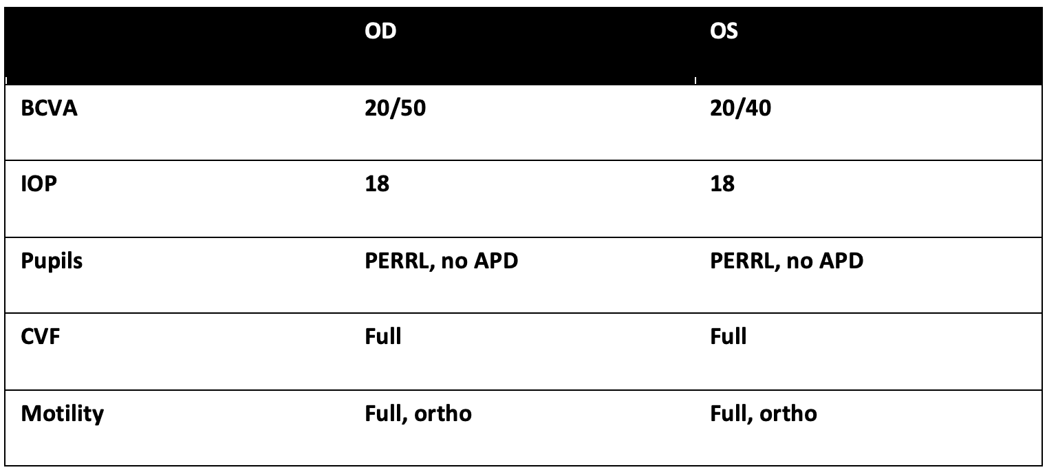
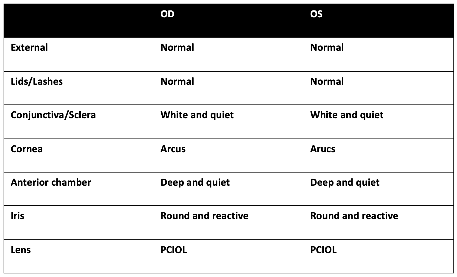
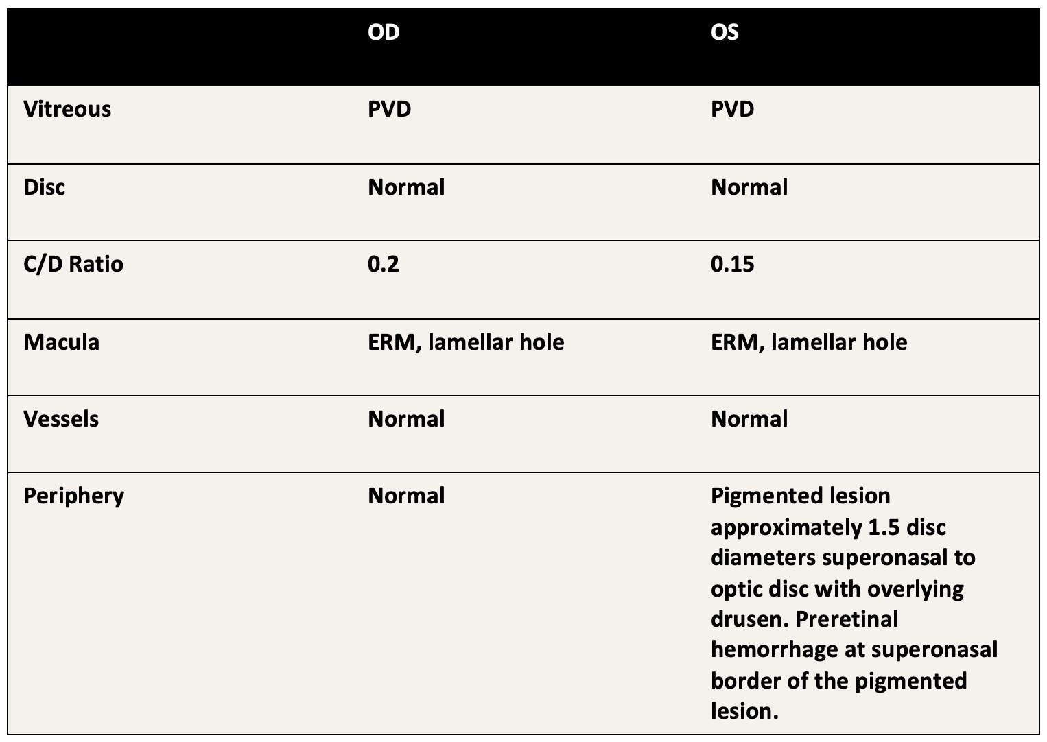
Exam
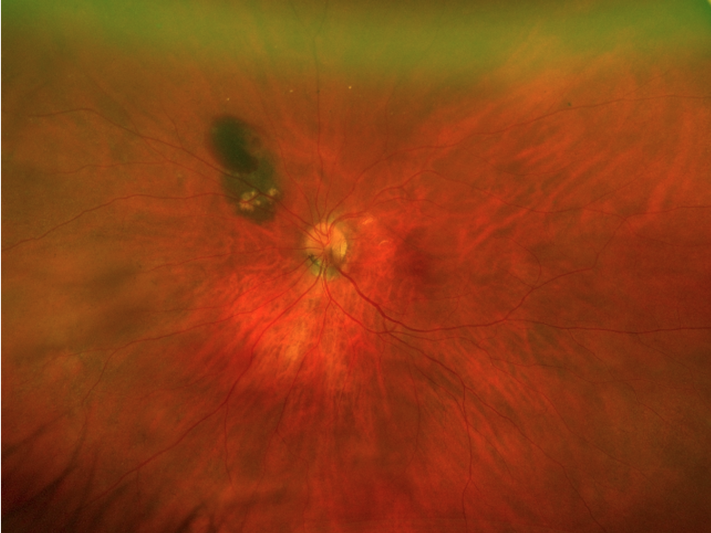
Fundus photograph of the left eye
Summary of Findings
- 90 year-old-man presents referred from optometry clinic for a pigmented lesion in the left eye with heme.
- Funduscopy: flat pigmented lesion approximately 1.5 disc diameters superonasal to optic disc with overlying drusen. Preretinal hemorrhage at superonasal border of the pigmented lesion.
- OCT (not pictured): OCT through lesion showing no height. OCT through hemorrhage with subretinal hyperreflective material.
Mechanism and differential diagnosis
- Choroidal nevus with associated choroidal neovascular membrane
- Peripheral Choroidal neovascular membrane
- Idiopathic polypoidal choroidal vasculopathy
- Vasoproliferative tumor
Treatment & clinical course
- The features of the pigmented lesion were most consistent with a choroidal nevus with an associated choroidal neovascular membrane.
- Given the lesion was far from the macula/fovea, observation was recommended.
- Close follow up over the next few months revealed resolution of the hemorrhage with no exudation into the macula.
- Diagnosis: choroidal nevus with an associated choroidal neovascular membrane
Discussion
- Choroidal nevi are a type of melanocytic tumor.
- Differentiating between choroidal nevi and melanomas are of the upmost importance. A helpful mnemonic in this determination is “To Find Small Ocular Melanomas Doing IMaging”: Thickness greater than 2mm on ultrasound, Fluid (subretinal) on OCT, Symptoms (vision changes), Orange pigment, Melanoma hollow on ultrasound, DIaMeter more than 5mm.1,2 The greater the number of items satisfied, the higher probability of the lesion being melanoma. In contrast, the presence of drusen and haloes suggest benign features.
- Choroidal neovascularization (CNV) associated with a choroidal nevus is a relatively rare complication which can cause vision changes and also distort the anatomy of the nevus thereby raising the suspicion for melanoma. As high as 1% of choroidal nevi can have an associated CNV. However, choroidal neovascularization is not thought to be a risk factor for malignant transformation of a choroidal nevus and often indicates chronicity.2-4 Intravitreal bevacizumab, photodynamic therapy, or even laser can be valid treatment options depending on the location of the nevus/CNV and the risk of vision threatening disease.4
- Multimodal imaging2,5 is helpful in determining the risk factor for choroidal melanoma in a choroidal lesion, especially in cases with confounding factors such as an associated CNV.
1. Shields CL, Furuta M, Berman EL, Zahler JD, Hoberman DM, Dinh DH, Mashayekhi A, Shields JA. Choroidal nevus transformation into melanoma: analysis of 2514 consecutive cases. Arch Ophthalmol. 2009 Aug;127(8):981-7.
2. Shields CL, Dalvin LA, Ancona-Lezama D, Yu MD, Di Nicola M, Williams BK Jr, Lucio-Alvarez JA, Ang SM, Maloney S, Welch RJ, Shields JA. CHOROIDAL NEVUS IMAGING FEATURES IN 3,806 CASES AND RISK FACTORS FOR TRANSFORMATION INTO MELANOMA IN 2,355 CASES: The 2020 Taylor R. Smith and Victor T. Curtin Lecture. Retina. 2019 Oct;39(10):1840-1851.
3. Callanan DG, Lewis ML, Byrne SF, Gass JD. Choroidal neovascularization associated with choroidal nevi. Arch Ophthalmol. 1993 Jun;111(6):789-94.
4. Chiang A, Bianciotto C, Maguire JI, Park CH, Baker PS, Shields JA, Shields CL. Intravitreal bevacizumab for choroidal neovascularization associated with choroidal nevus. Retina. 2012 Jan;32(1):60-7.
5. Pellegrini M, Corvi F, Say EAT, Shields CL, Staurenghi G. OPTICAL COHERENCE TOMOGRAPHY ANGIOGRAPHY FEATURES OF CHOROIDAL NEOVASCULARIZATION ASSOCIATED WITH CHOROIDAL NEVUS. Retina. 2018 Jul;38(7):1338-1346.