

CASE OF THE MONTH: April 2022
A Novel Retinopathy
History:
A 4-year-old girl presents for ophthalmic evaluation
Case
- Physical exam notable for short stature, cognitive delay, and microcephaly.
- She fixes and follows with full extraocular motility.
- Anterior segment examination notable for right iris coloboma.
- Cycloplegic refraction
- OD -9.00D
- OS -8.00D
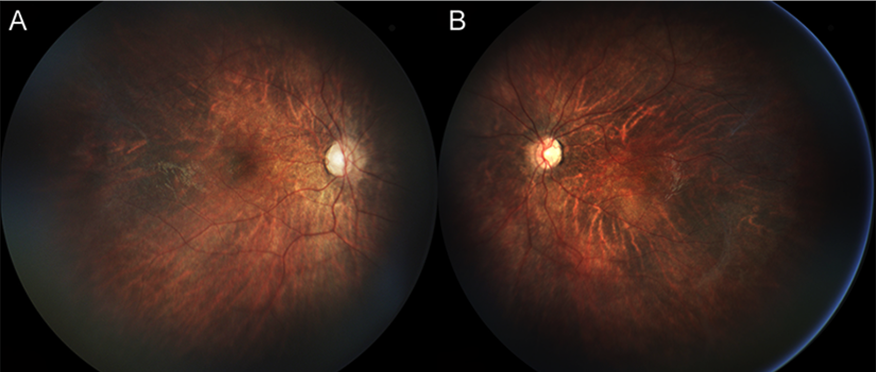
(A, B) Peripapillary atrophy and slightly attenuated vasculature with a truncated course
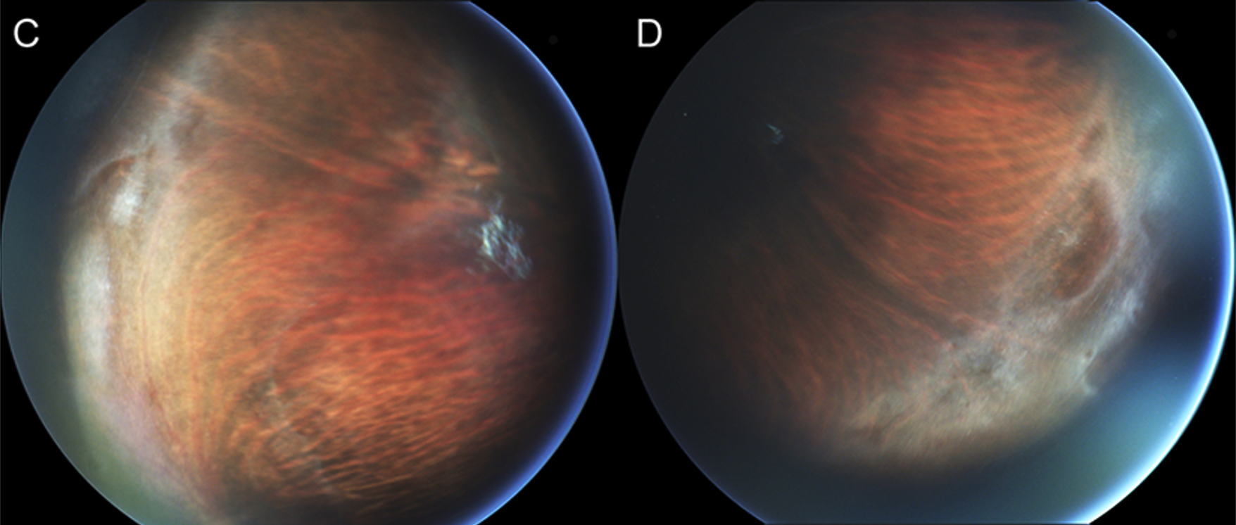
The peripheral retina was avascular with lattice degeneration
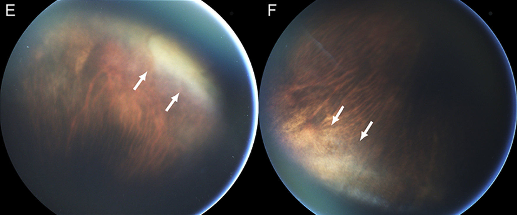
hyaloidal organization (white arrows).
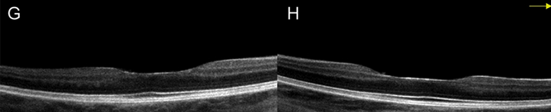
SDOCT of the macula (G,H) demonstrate flattening and widening of the foveal depression.
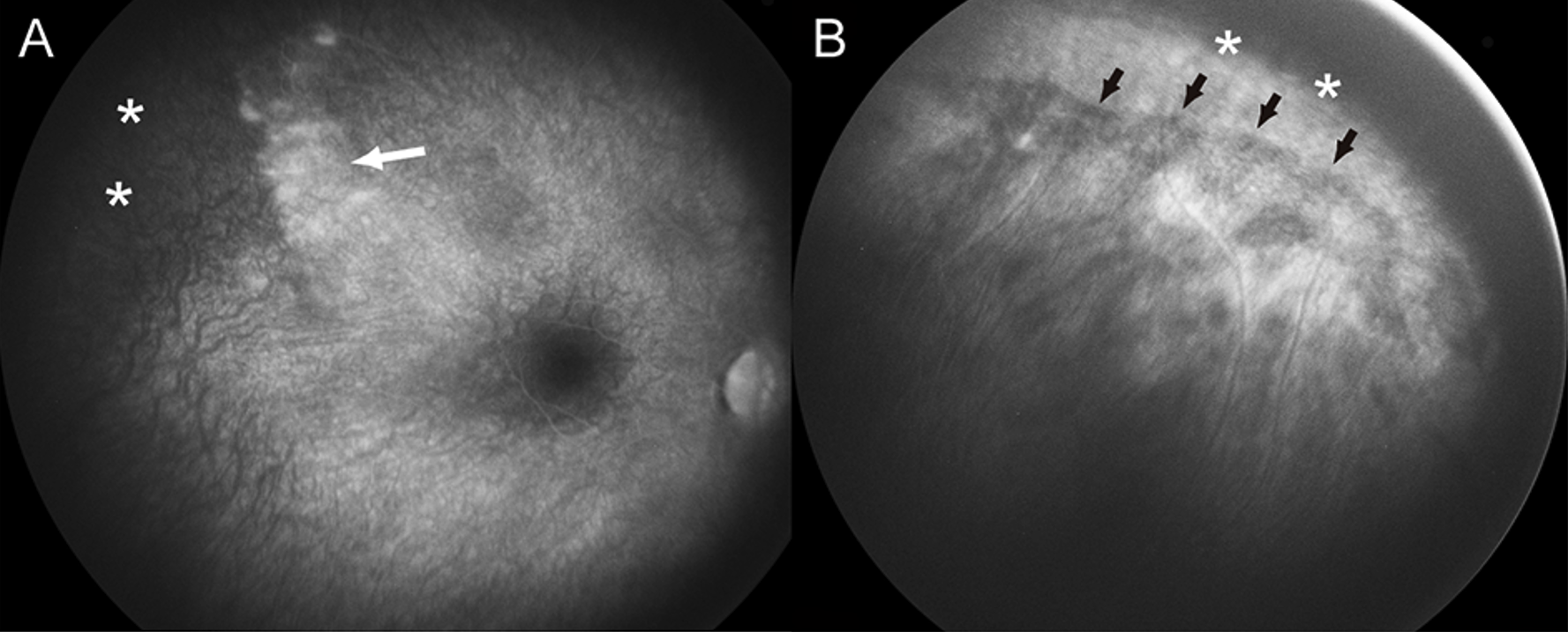
Widefield fluorescein angiography (FA), vascular pruning, and vascular arborization with leakage (Figure 2 ).
Genetic Testing...
- Our patient had a novel heterozygous mutation in exon 9 of the DYRK1A gene
- Dual-specificity tyrosine phosphorylation-regulated kinase 1A or DYRK1A is a proline directed kinase that plays a role in the development of the central nervous system.
- This mutation caused a frame shift leading to a premature stop codon and loss of function.
DYRK1A Syndrome
- Haploinsuffiency causes a cognitive deficiency syndrome associated with microcephaly, limb abnormalities, psychomotor delay, spinal and thoracic features, gastrointestinal features, cardiac disease, and renal features.1
- A survey of 145 patients with DYRK1A syndrome revealed ocular findings in 62%.
- 36% with refractive error (mix of myopic/amblyopic)
- 21% with strabismus
- 26% with enophthalmos
- 20% with optic nerve abnormalities
1. Our patient presented with iris coloboma, which has only been reported in rare instances, and abnormalities of the retinal vasculature, which have not been previously evaluated in detail.1,3
Similarity to FEVR
- The peripheral vasculature, especially at the vascular-avascular junction, resembled the vascular pruning and arborization seen in FEVR
- Relatively normal posterior vasculature and absence of supernumerary vasculature is slightly atypical
- Patient was not found to have any of the common genetic variants in FEVR (eg: FZD4, NDP, LRP5, TSPAN12, etc...)
- However, DYRK1A has been found to modulate the Wnt signaling pathway - important in the pathogenesis of FEVR
Normal 24 Gene Vitreoretinopathy Panel, Blueprint Genetic Inc, Seattle WA
Treatment
- Given the possibility of tractional retinal detachment which is seen in FEVR, laser photocoagulation was applied to the avascular retina in both eyes.
Conclusion
- We present the first detailed phenotypic analysis of the anomalous retinal vasculature in DYRK1A syndrome.
- Patients may benefit from clinical management that parallels that of FEVR due to the similarities:
- Widefield fluorescein angiography to identify the retinopathy,
- Laser photocoagulation to the avascular peripheral retina if there is angiographic leakage or neovascularization.
1. Méjécase C, Way CM, Owen N, Moosajee M. Ocular Phenotype Associated with DYRK1A Variants. Genes. 2021;12(2):234. doi:10.3390/genes12020234
2. Ernst J, Alabek ML, Eldib A, et al. Ocular findings of albinism in DYRK1A-related intellectual disability syndrome. Ophthalmic Genet. 2020;41(6):650-655. doi:10.1080/13816810.2020.1814349
3. Evers JMG, Laskowski RA, Bertolli M, et al. Structural analysis of pathogenic mutations in the DYRK1A gene in patients with developmental disorders. Hum Mol Genet. 2017;26(3):519-526. doi:10.1093/hmg/ddw409
4. Laguna A, Barallobre M-J, Marchena M-Á, et al. Triplication of DYRK1A causes retinal structural and functional alterations in Down syndrome. Hum Mol Genet. 2013;22(14):2775-2784. doi:10.1093/hmg/ddt125
5. Hong JY, Park J-I, Lee M, et al. Down’s-syndrome-related kinase Dyrk1A modulates the p120-catenin-Kaiso trajectory of the Wnt signaling pathway. J Cell Sci. 2012;125(Pt 3):561-569. doi:10.1242/jcs.086173
6. Gwack Y, Sharma S, Nardone J, et al. A genome-wide Drosophila RNAi screen identifies DYRK-family kinases as regulators of NFAT. Nature. 2006;441(7093):646-650. doi:10.1038/nature04631