

CASE OF THE MONTH: Q4 2026
90 year-old woman with progressively worsening vision for several years


Introduction
A 90-year-old woman presented with blurry vision and difficulty recognizing faces in both eyes that had been progressively worsening over several years. Patient notes she has a history of interstitial cystitis, for which she had been taking pentosan polysulfate sodium (PPS) for over 20 years. She reports no other significant past medical history, family history, or medication use.
On exam, she was noted to have a best corrected visual acuity of 20/40 in the right eye and 20/60 in the left eye. Intraocular pressure was within the normal range in both eyes. Anterior segment exam was unremarkable. Dilated fundoscopic exam was significant for pigment clumping and pigmentary changes in the macula of both eyes (Figure 1).
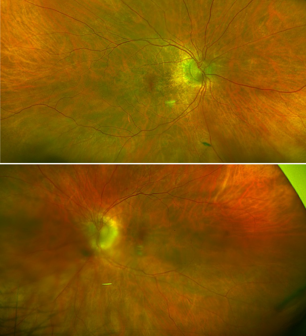
Figure 1. Widefield fundus photography (Top- Right Eye, Bottom- Left Eye) demonstrating pigment clumping and pigmentary changes within the macula in both eyes.
Work-Up
Ocular coherence tomography (OCT) showed areas of outer retinal irregularity, subretinal deposits, and focal areas of nodular RPE thickening in both eyes (Figure 2). Widefield fundus autofluorescence (FAF) demonstrated a dense speckled array of hyperautofluorescent and hypoautofluorescent spots within the macula of both eyes (Figure 3). The patient was subsequently diagnosed with PPS maculopathy based on clinical history, exam, and imaging findings. Macular pathologies that can have similar findings such as age-related macular degeneration, macular telangiectasia, pachychoroid spectrum disease, Stargardt’s disease, pattern dystrophy, cone/cone-rod dystrophy, and other inherited macular dystrophies were considered in the differential. Systemic diseases with associated retinopathy such as Bardet-Biedl and Alport syndrome were also considered. Other drug toxicities which can also affect the retinal pigment epithelium were also considered, but the patient did not have any other significant medication use. In the context of PPS use for over 20 years and a lack of significant family history or systemic findings, the other entities on the differential were considered unlikely.
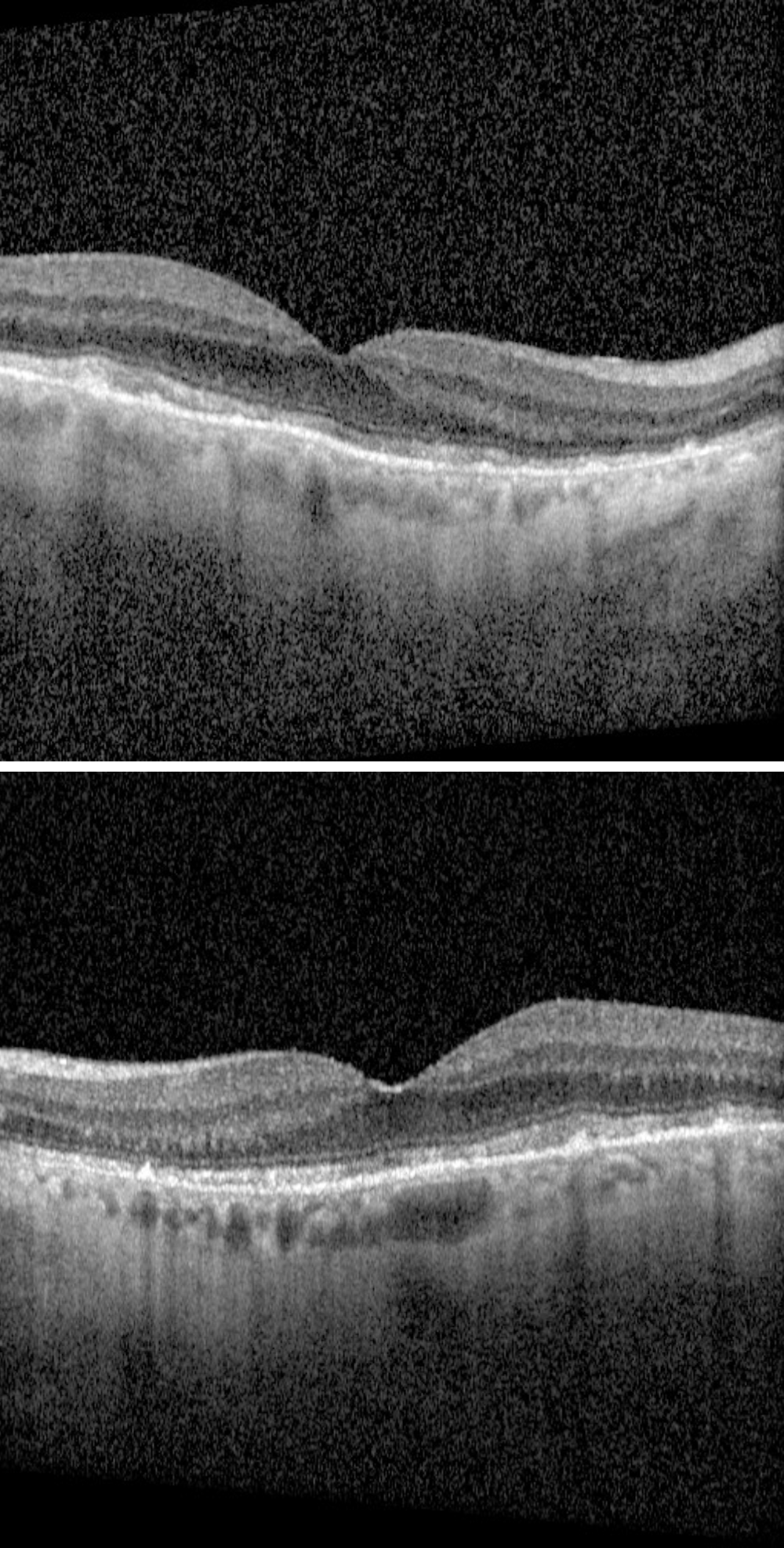
Figure 2. OCT imaging (Top-Right Eye, Bottom- Left Eye) demonstrating areas of outer retinal irregularity, subretinal deposits, and focal areas of nodular RPE thickening.
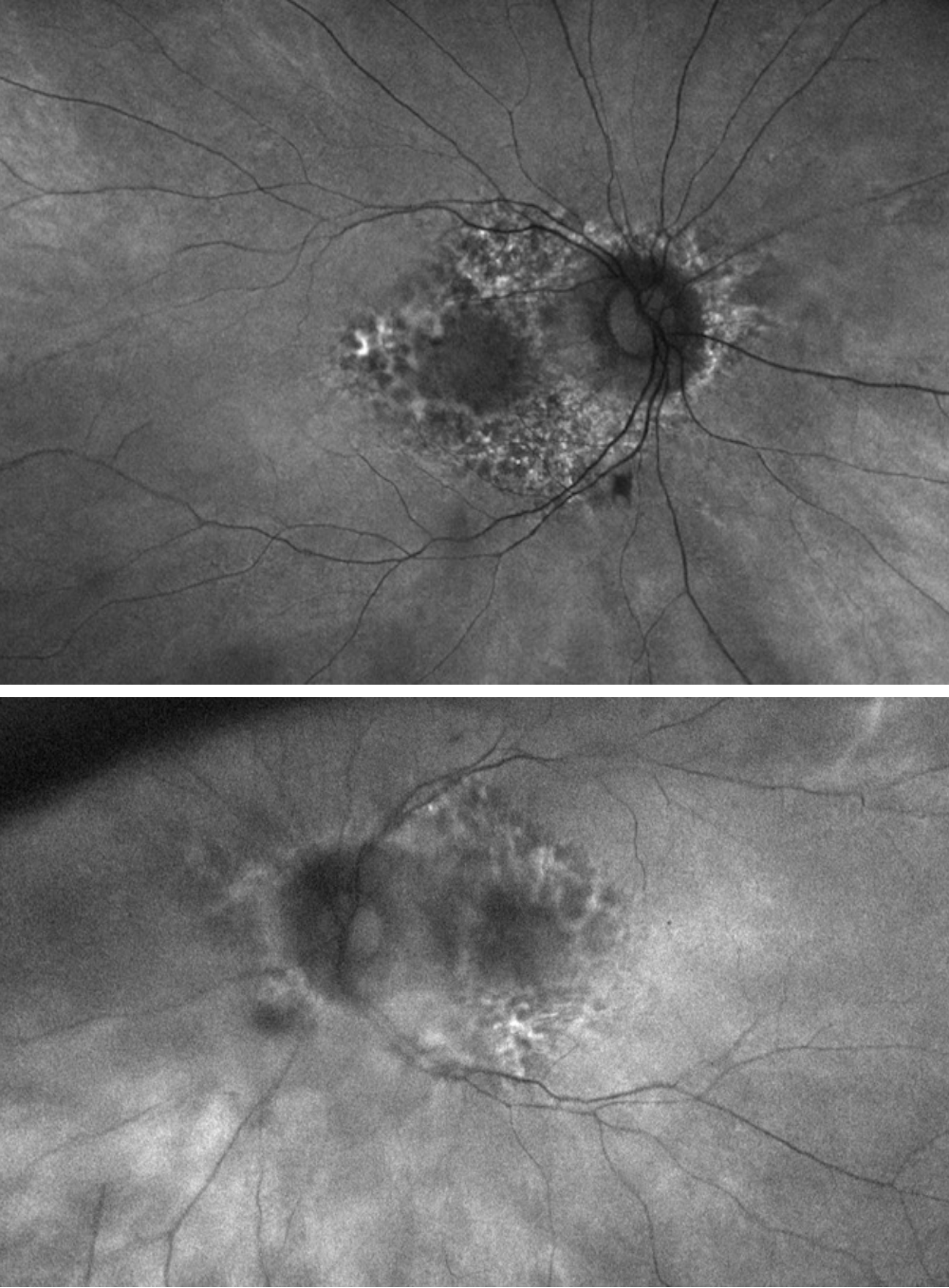
Figure 3. Fundus Autofluorescence imaging (Top-Right Eye, Bottom – Left Eye) demonstrating a dense speckled array of hyperautofluorescent and hypoautofluorescent spots.
Discussion
Pentosan polysulfate sodium (Trade Name- Elmiron) is an oral medication that was FDA approved in 1996 for treating interstitial cystitis, which is a syndrome characterized by urinary frequency, urinary urgency, and chronic pelvic pain without a identifiable cause. Interstitial cystitis affects more than 1 million patients in the United States and is much more common in women compared to men. PPS is thought to protect the bladder lining from irritants by replacing disrupted glycosaminoglycans along the bladder urothelium. Beginning in 2018, reports began to appear showing a maculopathy associated with long-term PPS use. Subsequent studies further substantiated the maculopathy as dose-dependent and characterized the clinical features.
Affected patients commonly experience blurred vision, prolonged dark adaptation, metamorphopsia, nyctalopia, scotomas, and difficulty reading, often with sparing of visual acuity. PPS retinopathy primarily manifests as a maculopathy but there are cases with involvement of the peripheral retina. PPS maculopathy can have RPE atrophy, outer retinal atrophy, cystoid macular edema, and choroidal neovascularization. Primary risk factors for development of PPS maculopathy is high cumulative PPS dose. Thus far, no other medical comorbidities or medications have been found to be independent risk factors for development of PPS maculopathy. Additionally, no genetic associations have been found that predispose patients to this maculopathy.
On exam, patients can present with hyperpigmented macular spots, yellow subretinal deposits, and patchy areas of RPE atrophy in advanced cases. However, some of these changes can be quite subtle to detect on fundoscopic exam. FAF imaging is often the most prominent showing a densely packed alternating array of hyperautofluorescent and hypoautofluorescent spots centered around the fovea in both eyes. A peripapillary halo of hypoautofluorescence can sometimes be seen in contrast to sparing of peripapillary FAF changes in hereditary maculopathies. OCT can show hyperreflective RPE nodular changes corresponding to macular pigment clumps, RPE atrophy, outer retinal irregularity, subretinal deposits, and retinal thinning.
The pathophysiology is still unknown, but it has been postulated that there may be direct toxicity to the RPE inhibiting processing of photoreceptor outer segments or disruption of the photoreceptor-RPE relationship. There no currently agreed upon severity scale for PPS maculopathy. Similarly, there is no consensus regarding screening guidelines for patients with PPS use, but it is important to screen patients at baseline and then yearly with multimodal imaging (FAF, OCT, and Fundus photography) to assess for any toxicity. Average duration of exposure to PPS is 10 years prior to maculopathy but there is significant variability in this, so yearly screening still makes sense. Furthermore, if there is already preexisting macular disease, PPS should generally be avoided or prescribed with significant caution and careful monitoring. Unfortunately, PPS maculopathy is thought to be irreversible and can still progress after cessation.
There are several entities which may appear like PPS toxicity and should be considered depending on the clinical scenario, patient history, and family history. Past medication and current medication use should be carefully assessed as drug toxicity from other medications such as hydroxychloroquine, iron chelators (e.g. deferoxamine), and alkylating chemotherapeutic agents (e.g. cisplatin) should be considered. Retinal dystrophies and degenerations such as AMD, Stargardt’s disease, pattern dystrophy, and cone-rod dystrophy among others should be considered. Systemic diseases with associated retinopathies such as Bardet-Biedl syndrome or Alport syndrome can also present similarly.
We explained to our patient that unfortunately PPS retinal toxicity is irreversible and stated that the medication should be stopped. We also explained there may still be progression of toxicity after stopping the medication. We discussed with the patient’s urologist that an alternative medication may be needed for treatment of interstitial cystitis.
References
1. Mukhopadhyay C, Boyce TM, Gehrs KM, et al. Age-Related Macular Degeneration Masquerade: A Review of Pentosan Polysulfate Maculopathy and Implications for Clinical Practice. Asia Pac J Ophthalmol (Phila). 2022;11:100–110.
2. Jain N, Li AL, Yu Y, VanderBeek BL. Association of macular disease with long-term use of pentosan polysulfate sodium: findings from a US cohort. British Journal of Ophthalmology. 2020;104:1093–1097.
3. Characteristics of Pentosan Polysulfate Sodium–Associated Maculopathy and Similarities With Other Maculopathies Commonly Managed in a Retina Practice. https://journals.sagepub.com/doi/epub/10.1177/24741264211020259 ; Accessed 17.06.26.
4. Hanno PM, Erickson D, Moldwin R, et al. Diagnosis and treatment of interstitial cystitis/bladder pain syndrome: AUA guideline amendment. J Urol. 2015;193:1545–1553.
5. Pearce WA, Chen R, Jain N. Pigmentary Maculopathy Associated with Chronic Exposure to Pentosan Polysulfate Sodium. Ophthalmology. 2018;125:1793–1802.
6. Kalaw FGP, Arias JD, Walker E, et al. REFINING THE RETINAL PHENOTYPE OF PATIENTS WITH SUSPECTED PENTOSAN POLYSULFATE SODIUM RETINOPATHY. RETINA. 2023;43:1472